In plain English
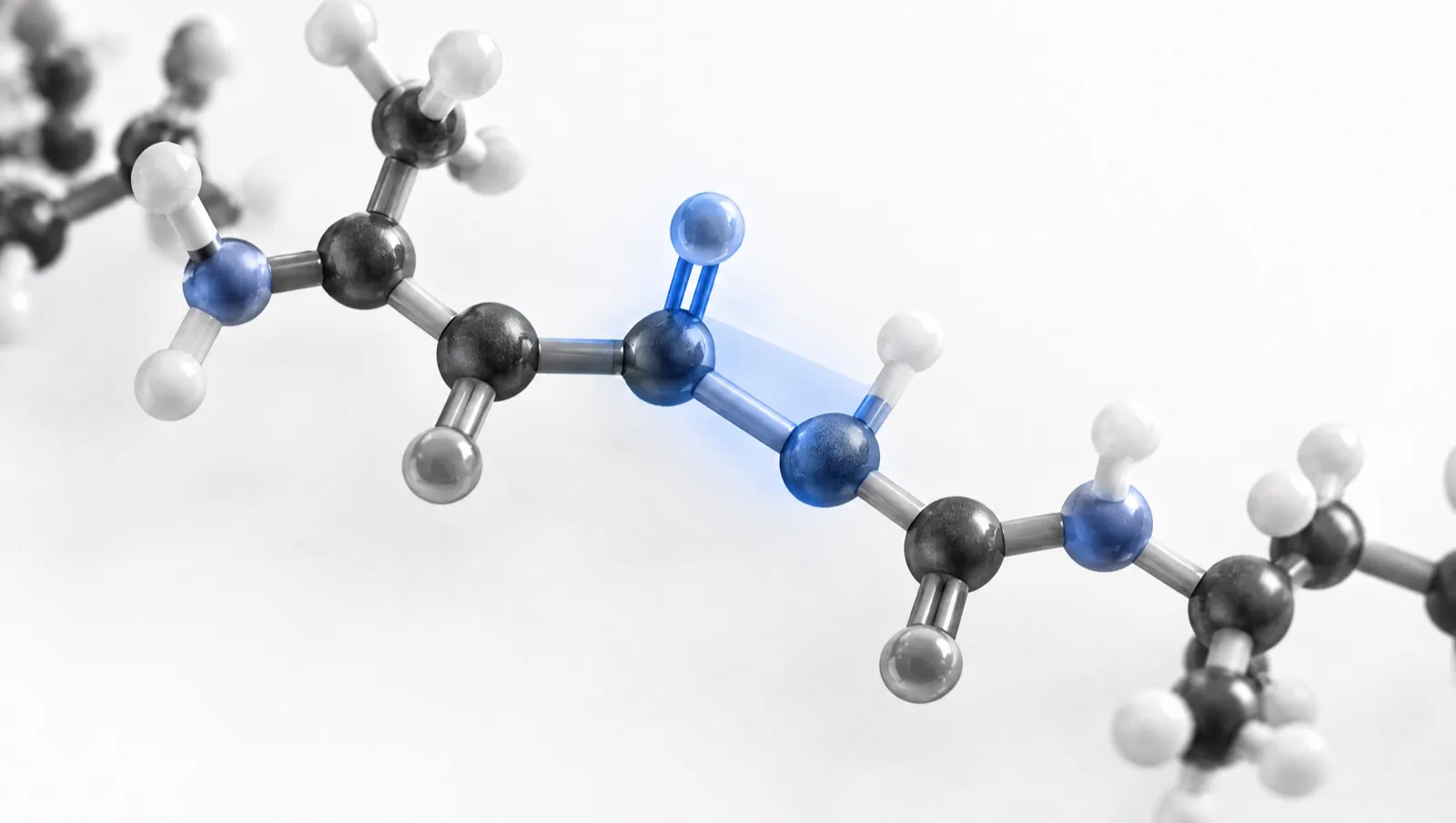
A peptide bond is the link between two amino acids in a chain. It forms when one amino acid’s acid end joins to the next one’s amino end. A small molecule of water is released when this happens [1].
The bond is stiff and flat. It behaves partly like a double bond, so it does not spin freely [2]. Most peptide bonds sit in one layout, called “trans”. Bonds just before the amino acid proline are the main exception: they flip between two layouts more easily [3].
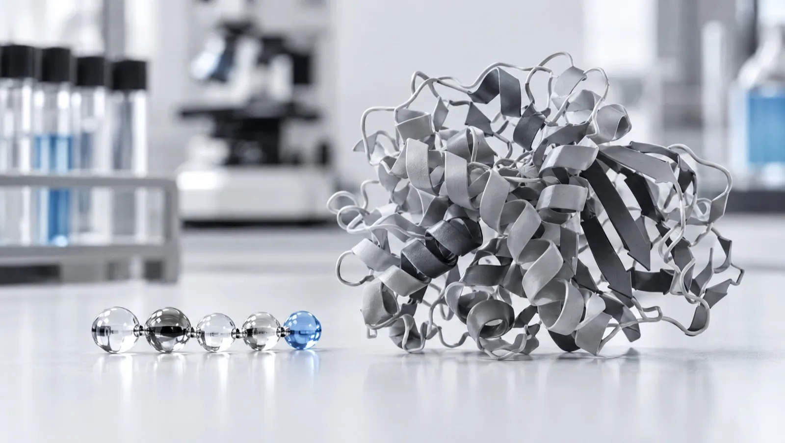
Because each link is flat, a chain can only bend at certain points. That limits the shapes a chain can take. Long chains can form regular shapes such as coils (α-helices) and flat strands (β-strands) [4]. Many short peptides do not hold one fixed shape in solution [5, 6].
The short version
- A peptide bond is an amide bond between amino acids [1].
- It is flat and usually trans [2, 3].
- Chains fold through rotation at the bonds beside it [7].
Key facts
| Property | Detail |
|---|---|
| Chemical type | Amide bond (–CO–NH–) between the carboxyl group of one amino acid and the amino group of the next [1] |
| Formation | Condensation; the elements of water are removed [1] |
| Geometry | Generally planar, because of partial double-bond character [2] |
| Usual arrangement | Trans, with the backbone torsion angle omega (ω) near 180° [2] |
| Main exception | Bonds preceding proline, where cis and trans are close in energy [3] |
| Backbone angles that vary | Phi (φ) and psi (ψ), plotted on a Ramachandran plot [7] |
| Breakdown | Amide bonds can be hydrolysed, including by enzymes [5] |
How a peptide bond forms
The IUPAC-IUB naming rules define a peptide as any compound produced by amide formation between a carboxyl group of one amino acid and an amino group of another. These amide bonds may be called peptide bonds [1].
When the bond forms, the elements of water are removed. What remains of each amino acid is called an amino-acid residue [1]. A dipeptide has one peptide bond, a tripeptide has two, and so on.
The rules separate two kinds of amide link [1]:
- Eupeptide bonds join the backbone carboxyl (C-1) of one amino acid to the α-amino group (N-2) of the next. This is the normal peptide bond.
- Isopeptide bonds use a side-chain group instead. Glutathione is an example: its glutamic acid is joined through its side-chain (γ) carboxyl group, so it is written as γ-glutamylcysteinylglycine [1, 8].
The chain that results has two distinct ends. The residue with the free (or at least not peptide-bonded) amino group is the N-terminus. The residue with the free carboxyl group is the C-terminus [1]. That direction matters for writing sequences. See How to Read a Peptide Sequence.
Why the peptide bond is flat
Peptide groups are generally treated as planar because the peptide bond has “rigid” partial double-bond character [2]. In practice, this means the atoms of each peptide group (C=O and N–H) and the two α-carbons they connect lie close to a single plane [2].
The angle that describes rotation about the peptide bond itself is called omega (ω). For the usual trans form, ω is close to 180° [2].
Planarity is a strong tendency, not an absolute rule. A study of ultra-high-resolution protein crystal structures found cases where ω deviated by more than 10° from 180°, showing measurable non-planarity in some peptide groups [2].
Trans and cis peptide bonds, and why proline is special
Because the peptide bond does not rotate freely, it can sit in two arrangements:
- Trans. The two α-carbons are on opposite sides of the bond. For ordinary peptide bonds in short peptides, the trans form is strongly preferred under almost all conditions [9].
- Cis. The two α-carbons are on the same side.
Proline is the main exception. Proline’s side chain is a ring that closes back onto its own backbone nitrogen, making it a secondary amine [1, 17]. For peptide bonds immediately preceding proline, cis and trans states are close in energy, so cis bonds are most commonly found there [3]. Switching between cis and trans still has to cross a high energy barrier, so it is slow [3].
Structural surveys of the Protein Data Bank highlight proline’s unique role in determining local conformation, including the relative occurrence of cis and trans bonds before proline residues [10]. In folding studies, slow cis–trans isomerisation can become the rate-determining step. One experiment on a ribonuclease variant found that re-isomerisation of a single non-proline cis bond slowed refolding about 1,000-fold [9].
Backbone angles and the Ramachandran plot
If the peptide bond is fixed, where does a chain bend? It bends at the two single bonds on either side of each α-carbon. Their rotation angles are called phi (φ) and psi (ψ).
In 1963, Ramachandran and colleagues analysed the stereochemistry of polypeptide chain configurations [11]. The resulting Ramachandran plot maps protein structure by plotting pairs of backbone dihedral angles on a two-dimensional chart [7]. Residues are not spread evenly across the plot. They cluster in a limited number of regions, including one associated with the α-helix [7].
Levels of peptide and protein structure
| Level | What it describes | Example |
|---|---|---|
| Primary | The sequence of residues, from N-terminus to C-terminus [1] | Oxytocin: Cys-Tyr-Ile-Gln-Asn-Cys-Pro-Leu-Gly-NH₂ [12] |
| Secondary | Regular local patterns formed by backbone hydrogen bonds; mainly α-helices and β-strands, which can form β-sheets [4] | Helical segments in glucagon [6] |
| Tertiary | The overall three-dimensional fold of a chain [4] | Folded proteins |
| Covalent cross-links | Disulfide bonds between cysteine side chains that lock parts of a chain, or separate chains, together [12, 13] | Oxytocin’s 1–6 disulfide ring; insulin’s A and B chains [12, 13] |
Secondary structure
Secondary structures are highly regular local substructures formed by hydrogen bonding along the backbone. The two main types are α-helices and β-strands, and β-strands can line up to form β-sheets [4]. In 1951, Pauling, Corey and Branson described two hydrogen-bonded helical configurations of the polypeptide chain, a landmark in understanding these patterns [14].
Circular dichroism (CD) spectroscopy is used to estimate secondary-structure content in peptides and proteins [4].
Do short peptides have a fixed structure?
Often they do not. A 2022 review notes that natural peptides lack the stability conferred by secondary or tertiary structures, which leaves their amide bonds open to hydrolysis by enzymes [5]. Glucagon, a 29-amino-acid hormone, illustrates this. A CD and NMR study of glucagon products classed about half of the structure as unordered and only about 14–17% as α-helix [6]. The same paper notes that glucagon can convert into a different, β-sheet-rich structure under some conditions [6]. That kind of change is the starting point for Peptide Aggregation.
Chemists sometimes stabilise a chosen shape on purpose. Cyclisation and side-chain cross-links can pre-organise a peptide and stabilise helices or sheets [5]. See Peptide Modifications Explained.
Why structure matters for laboratory work
- Identity is more than mass. Two peptides with the same composition but a different order of residues, or with an L residue swapped for its D form, have the same mass. That is why identity testing and purity testing are separate questions. See How to Read a Peptide COA.
- Disulfide bonds. Peptides with cysteine pairs, such as oxytocin, need the correct disulfide link to match the reference structure [12].
- Stability. Peptide bonds can be hydrolysed, and short peptides with little fixed structure are especially exposed [5]. See Peptide Storage and Stability.
What this means in Australia
Peptide products are regulated as therapeutic goods. The TGA states that peptide products, which it describes as short chains of amino acids, are regulated as therapeutic goods under the Therapeutic Goods Act 1989. Products not included on the Australian Register of Therapeutic Goods (ARTG) are unapproved [15].
Status attaches to the specific substance. The Poisons Standard (October 2026) names individual peptides, sometimes with an identifier. For example, it names CJC-1295 (CAS 863288-34-0) in Schedule 4 and Appendix D, clause 5 [16]. Schedule 4 means prescription-only. Appendix D, clause 5 means possessing the substance without authority is illegal. Some peptides are captured through class entries instead, and whether a class entry captures a specific compound is a legal question.
“Research use only” labels. The TGA has stated that a “research use only” disclaimer does not make supply lawful, and does not change a product’s regulatory status, permit importation, or remove advertising or supply obligations [15].
This is general information, not legal advice. Laws change; check the current Poisons Standard and TGA guidance.
Frequently asked questions
What is a peptide bond?
It is the amide bond formed between the carboxyl group of one amino acid and the amino group of another, with the loss of water [1].
Is a peptide bond the same as an amide bond?
Yes. A peptide bond is an amide bond in a peptide. The term usually refers to backbone links, while links through side chains are called isopeptide bonds [1].
Why can’t a peptide bond rotate freely?
It has partial double-bond character, which holds the peptide group roughly flat [2].
Why is proline different?
For bonds just before proline, the cis and trans forms are close in energy, so cis bonds occur much more often there than elsewhere [3].
Do peptides have secondary structure?
Some do, but many short peptides are partly or mostly unordered in solution. Proteins usually have stable helices and sheets [4, 5, 6].
References
- IUPAC-IUB Joint Commission on Biochemical Nomenclature (JCBN). Nomenclature and symbolism for amino acids and peptides. Recommendations 1983. Biochem J. 1984;219(2):345–373. doi:10.1042/bj2190345. PMID: 6743224. Web version: iupac.qmul.ac.uk/AminoAcid. Accessed 11 October 2026.
- Dasgupta AK, Majumdar R, Bhattacharyya D. Characterization of non-planar peptide groups in protein crystal structures. Indian J Biochem Biophys. 2004;41(5):233–240. PMID: 22900279
- Chen J, Edwards SA, Gräter F, Baldauf C. On the cis to trans isomerization of prolyl-peptide bonds under tension. J Phys Chem B. 2012;116(31):9346–9351. doi:10.1021/jp3042846. PMID: 22770126
- Kuril AK, Vashi A, Subbappa PK. A comprehensive guide for secondary structure and tertiary structure determination in peptides and proteins by circular dichroism spectrometer. J Pept Sci. 2025;31(1):e3648. doi:10.1002/psc.3648. PMID: 39135381
- Wang L, Wang N, Zhang W, et al. Therapeutic peptides: current applications and future directions. Signal Transduct Target Ther. 2022;7(1):48. doi:10.1038/s41392-022-00904-4. PMID: 35165272
- Bao Z, Cheng YC, Wei JJ, Luo MZ, Zhang JY. Secondary structure characterization of glucagon products by circular dichroism and nuclear magnetic resonance spectroscopy. Molecules. 2022;27(22):7805. doi:10.3390/molecules27227805. PMID: 36431905
- Park SW, Lee BH, Song SH, Kim MK. Revisiting the Ramachandran plot based on statistical analysis of static and dynamic characteristics of protein structures. J Struct Biol. 2023;215(1):107939. doi:10.1016/j.jsb.2023.107939. PMID: 36707040
- National Center for Biotechnology Information. PubChem Compound Summary for CID 124886, Glutathione. pubchem.ncbi.nlm.nih.gov/compound/124886. Accessed 11 October 2026.
- Odefey C, Mayr LM, Schmid FX. Non-prolyl cis-trans peptide bond isomerization as a rate-determining step in protein unfolding and refolding. J Mol Biol. 1995;245(1):69–78. doi:10.1016/s0022-2836(95)80039-5. PMID: 7823321
- MacArthur MW, Thornton JM. Influence of proline residues on protein conformation. J Mol Biol. 1991;218(2):397–412. doi:10.1016/0022-2836(91)90721-h. PMID: 2010917
- Ramachandran GN, Ramakrishnan C, Sasisekharan V. Stereochemistry of polypeptide chain configurations. J Mol Biol. 1963;7:95–99. doi:10.1016/s0022-2836(63)80023-6. PMID: 13990617
- National Center for Biotechnology Information. PubChem Compound Summary for CID 439302, Oxytocin. pubchem.ncbi.nlm.nih.gov/compound/439302. Accessed 11 October 2026.
- UniProt Consortium. UniProtKB entry P01308, Insulin (human). uniprot.org/uniprotkb/P01308/entry. Accessed 11 October 2026.
- Pauling L, Corey RB, Branson HR. The structure of proteins; two hydrogen-bonded helical configurations of the polypeptide chain. Proc Natl Acad Sci U S A. 1951;37(4):205–211. doi:10.1073/pnas.37.4.205. PMID: 14816373
- Therapeutic Goods Administration. Understanding your responsibilities when importing, compounding and supplying unapproved peptide products. Safety advisory, 13 April 2026. tga.gov.au. Accessed 11 October 2026.
- Therapeutic Goods (Poisons Standard—October 2026) Instrument 2026 (F2026L01327). Federal Register of Legislation. legislation.gov.au/F2026L01327/latest/text. Accessed 11 October 2026.
- Lopez MJ, Mohiuddin SS. Biochemistry, Essential Amino Acids (Archived). In: StatPearls. Treasure Island (FL): StatPearls Publishing; 2024. PMID: 32496725
Important information and disclaimer
General information only. This article is published for general educational and informational purposes. It summarises published scientific literature and public regulatory information as at the “last reviewed” date shown above. It is not medical, health, pharmaceutical, veterinary or legal advice, and it must not be relied on as a substitute for advice from a qualified professional about your circumstances.
Not for human or animal use. Nothing in this article recommends, endorses or describes the use of any substance in humans or animals. Findings from cell, animal or early human studies cannot be assumed to apply to people. The compounds discussed have not been evaluated by the Therapeutic Goods Administration (TGA) for safety, quality or efficacy unless the article expressly states otherwise. Any research materials offered by Iconic Peptides are supplied solely for in vitro and laboratory research.
No therapeutic claims. Iconic Peptides makes no claim that any product treats, cures, prevents, alleviates or diagnoses any disease, condition or symptom. References to published studies, clinical trials or medicines approved by regulators are factual descriptions of the scientific and regulatory record. They are not representations about any product we supply.
Regulatory status. Many peptides are scheduled substances under the Poisons Standard. Some are prescription-only (Schedule 4), and some may not be possessed without authority (Appendix D). The TGA has stated that a “research use only” label does not make supply lawful, change a product’s regulatory status, or remove advertising or supply obligations. State and territory laws may also apply. You are responsible for knowing and complying with all laws that apply to you. Regulatory information in this article may change after the review date.
Accuracy and third-party sources. We take care to cite and verify every source, but we do not warrant that this article is complete, current or error-free. Citing a study or organisation does not mean that its authors or that organisation endorse Iconic Peptides.
Limitation of liability. To the maximum extent permitted by law, Iconic Peptides, its owners, employees and contributors exclude all liability for any loss, damage, injury or expense, whether direct, indirect or consequential, arising from use of or reliance on this article or any misuse of any product. Nothing in this disclaimer excludes, restricts or modifies any right or remedy under the Australian Consumer Law that cannot lawfully be excluded.
Age restriction. This website is intended for adults aged 18 and over.