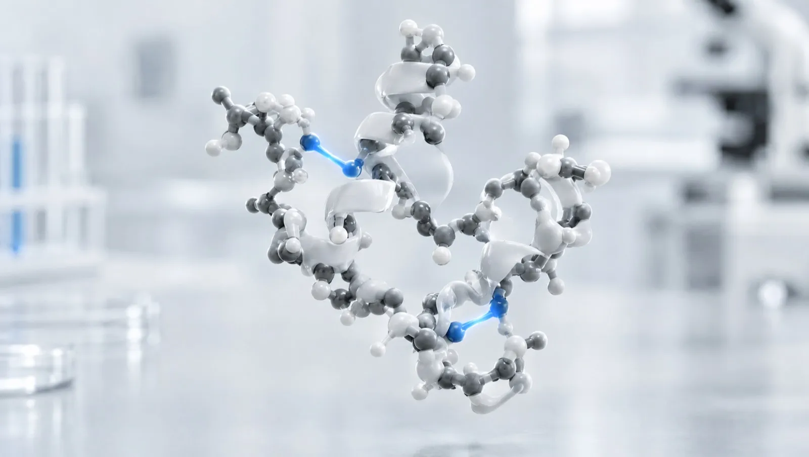
In plain English
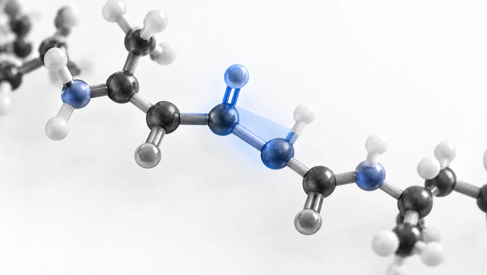
Cysteine is the amino acid with a sulfur atom at the end of its side chain, carried as a thiol group (–SH). Two thiols can be joined by removing one hydrogen from each, leaving a sulfur–sulfur bridge. The joined pair has its own name: cystine [1, 2].
The bridge acts like a staple. It holds two parts of a chain, or two separate chains, together. Insulin is the classic example: it has three such bridges, two joining its two chains and one inside the shorter chain [16].
Two things follow for laboratory work. First, because two hydrogen atoms are lost, the bridged molecule is about 2 Da lighter than the unbridged one, which a mass spectrometer can see [11]. Second, when a peptide has four, six or more cysteines, they can pair up in several different patterns. Only one pattern is usually wanted, and the wrong ones have the same mass as the right one [3].
The short version
- A disulfide bond joins two cysteine side chains [1, 2].
- Forming one makes the molecule about 2 Da lighter [11].
- With several cysteines, several bridge patterns are possible [3].
- Bridges rearrange fastest in alkaline solution [5].
Key facts
| Property | Detail |
|---|---|
| The residue | L-cysteine, C₃H₇NO₂S, about 121.16 g/mol, with a free thiol side chain [1] |
| The linked pair | L-cystine, C₆H₁₂N₂O₄S₂, about 240.3 g/mol [2] |
| Mass change per bridge | −2.016 Da (two hydrogen atoms removed) [11, 17] |
| Reactive species in exchange | The thiolate (–S⁻), about 10¹⁰ times more reactive than the protonated thiol [5] |
| Possible isomers | (2n)!/(2ⁿn!): 3 for two bridges, 15 for three, 105 for four [3] |
| Common free-thiol assay | Ellman’s reagent (DTNB), read at 412 nm [5, 7, 8] |
| Common reductants | Dithiothreitol (DTT), tris(2-carboxyethyl)phosphine (TCEP) [5, 9, 10] |
From cysteine to cystine
A thiol group is a weak acid. Only its deprotonated form, the thiolate, is an effective nucleophile: the protonated forms of simple alkyl thiols react some 10¹⁰-fold more slowly than their thiolates [5]. Biological thiols span roughly pKa 3 to 11, an eight-order-of-magnitude span in how much thiolate is present at a given pH [5].
That single fact explains most of the practical behaviour of disulfide chemistry. Oxidation, exchange and scrambling all run through the thiolate, so all accelerate as pH rises. For a thiol–disulfide exchange the observed rate constant follows
kobs = k / (1 + 10^(pKa − pH))
so the rate is half its maximum at the pKa and falls to one ten-thousandth of the maximum four pH units below it [5]. The mechanism is a thiolate attacking one sulfur of the target disulfide through a transition state in which the three sulfur atoms lie in a line, giving a mixed disulfide and releasing a new thiolate as the leaving group [5, 6].
The 2 Da rule, and a worked example
Forming a disulfide removes one hydrogen from each of two thiols. Two hydrogen atoms is 2.0157 Da, so the oxidised (bridged) form is about 2 Da lighter than the reduced form, and reducing a peptide with n disulfides adds about 2n Da.
Oxytocin illustrates this: a nonapeptide with a single 1–6 disulfide. PubChem gives the cyclic molecule as C₄₃H₆₆N₁₂O₁₂S₂, monoisotopic mass 1006.436 Da [17]. The reduced dithiol form is C₄₃H₆₈N₁₂O₁₂S₂:
| Form | Monoisotopic mass | Singly protonated ion |
|---|---|---|
| Oxidised (1–6 disulfide) | 1006.44 Da | m/z ≈ 1007.44 |
| Reduced (two free thiols) | 1008.45 Da | m/z ≈ 1009.46 |
This is why identity testing of a cystine-containing peptide should state which form was measured. The general strategy for assigning bridges uses exactly this arithmetic: masses of a non-reduced digest are compared with masses of a reduced aliquot of the same digest [11]. See Reading a Peptide Mass Spectrum.
How disulfides are formed: oxidative folding
The simplest route is to deprotect every cysteine and let the bridges form in one step. Published conditions for disulfide-rich peptides, summarised in a 2014 review of conotoxin chemistry, include [3]:
- Air oxidation in mildly alkaline buffer, typically pH 7.5–8.5, at high dilution (around 200–500 µM) to favour intramolecular over intermolecular bridges.
- A thiol/disulfide redox buffer, for example reduced and oxidised glutathione with the peptide in a ratio of about 100:10:1, which lets wrongly formed bridges reshuffle towards the most stable arrangement.
- Denaturants such as 0.5–3.0 M urea or 0.1–1.5 M guanidine hydrochloride, where aggregation is a problem.
- DMSO-promoted oxidation, which works from about pH 1 to pH 8, is faster in acidic media, and also helps disperse aggregates and dissolve hydrophobic sequences; 10% or even 50% DMSO in 1 M HCl gives high yields, although removing the DMSO afterwards can be awkward.
- Iodine, a strong oxidant used mainly for directed bridge formation (below).
The trade-off is plain: the more reactive the oxidant, the more side reactions, particularly at methionine, tryptophan and tyrosine [3].
Why random oxidation is not enough
Folding a disulfide-rich peptide involves oxidation, reduction and isomerisation (scrambling) at once, and the number of possible pairings grows fast. For n disulfides there are (2n)!/(2ⁿn!) isomers: 3 for two bridges, 15 for three, and 105 for four [3]. Usually only one is the intended structure, and the non-native isomers share its mass and often its retention time [3]. Random oxidation works when the sequence encodes a clearly most-stable fold, and fails when it does not — for example once non-natural residues or other modifications are built into the design [3].
Directed (regioselective) bridge formation
The alternative is to form bridges one pair at a time, using cysteine protecting groups that come off under different conditions. More than sixty individual protecting groups have been reported for the cysteine thiol, and the choice of orthogonal set is what makes complex multi-disulfide syntheses possible [4].
| Group | Removed by | Typical role |
|---|---|---|
| Trt (trityl) | Acid, during the usual TFA cleavage | Gives the free thiols oxidised first |
| Acm (acetamidomethyl) | Iodine or thallium trifluoroacetate, forming the bridge directly; or mercury salts to give free thiols | The workhorse for a second, directed bridge [3] |
| Mob, Meb, tBu | Acid, or acid/DMSO conditions at a chosen temperature | A third orthogonal pair in three-bridge syntheses [3] |
| Fm (fluorenylmethyl) | Base, for example piperidine | Allows an on-resin bridge before cleavage [3] |
A common off-resin sequence is: cleave the peptide so that every protecting group except Acm is removed, oxidise the now-free cysteine pair with a mild oxidant to make the first bridge, then form the second bridge directly with iodine on the Acm-protected pair [3]. A third orthogonal pair (for example S-Mob alongside S-Acm and S-Trt) allows all three bridges of a three-disulfide peptide to be closed consecutively [3].
Two cautions come with this. Conditions must avoid breaking or scrambling bridges already in place, which means avoiding alkaline conditions, free thiols, other nucleophiles and long reaction times [3]. And the Acm-removal step needs care, because iodine and thallium reagents can oxidise methionine, histidine, tryptophan and tyrosine [3].
Scrambling, and how to slow it down
Once formed, a disulfide is not inert. It can exchange with any thiolate that reaches it, which means samples can rearrange between the bench and the instrument.
| Condition | Effect |
|---|---|
| Alkaline pH | Accelerates exchange, because more thiol is present as thiolate [5] |
| Acidic pH | Slows exchange sharply; acidification is the usual way to quench a redox reaction [5] |
| Trace metal ions | Cu²⁺ > Fe³⁺ > Ni²⁺ ≫ Co²⁺ stimulate oxidation of thiols by dissolved oxygen; chelators such as EDTA attenuate but do not abolish it [5] |
| Dissolved oxygen | A source of artefactual disulfide; purging with nitrogen or argon is recommended where it matters [5] |
| Free thiols elsewhere in the sample | Provide the nucleophile that starts the exchange [5, 6] |
Degradation studies show the pH effect concretely. In an accelerated study of oxytocin formulations at 40–80 °C, loss of intact peptide was fastest at pH 9.0, then pH 7.0, then pH 2.0, and slowest at pH 4.5; at pH 4.5–9.0 the identified products included tri- and tetrasulfide forms, disulfide-linked and dityrosine-linked dimers, β-elimination products and larger aggregates [15]. A review of thiols and disulfides in protein stability describes the same two families of consequence: chemical degradation through oxidation and β-elimination, and physical degradation through aggregation and precipitation [14]. See Peptide Storage and Stability.
Measuring free thiols: Ellman’s reagent
Thiols are measured directly; disulfides are usually measured indirectly, by reducing them and measuring the thiols that appear [5].
The classical reagent is 5,5′-dithiobis(2-nitrobenzoic acid), DTNB, introduced for tissue sulfhydryl groups in 1959 [7] and re-examined in 1979 [8]. DTNB carries a highly oxidising disulfide; a free thiol reduces it stoichiometrically, releasing one molecule of the strongly coloured 5-thio-2-nitrobenzoate (TNB) per thiol reacted [5].
A review of thiol and disulfide quantification gives the TNB molar absorption coefficient as 14,100 M⁻¹ cm⁻¹ at 412 nm and pH 7.3, dropping to 13,700 M⁻¹ cm⁻¹ in 6 M guanidinium chloride, and notes that absorbance falls steeply below neutral pH as the orange thiolate becomes protonated [5].
Worked example
A sample gives A₄₁₂ = 0.282 in a 1 cm cell after reaction with DTNB, against a reagent blank.
- c = A / (ε × l) = 0.282 / (14,100 M⁻¹ cm⁻¹ × 1 cm) = 2.0 × 10⁻⁵ M = 20 µM thiol
- If the peptide is present at 20 µM and should contain one disulfide and no free cysteine, 20 µM of thiol is one free thiol per molecule — a signal that the bridge has not formed, or has been reduced.
Two warnings apply. DTNB hydrolyses at elevated temperature and above pH 7, which generates a sulfenate and a thiolate and so inflates blanks [5]. And a reagent whose absorbance is pH-independent, such as 4,4′-dithiodipyridine (ε 21,000 M⁻¹ cm⁻¹ at 324 nm, usable from about pH 3 to 7), may be preferable when work has to be done at low pH to suppress exchange [5].
Reducing disulfides on purpose
| Reductant | Character | Notes |
|---|---|---|
| DTT | Dithiol; low redox potential because exchange closes a six-membered ring | Introduced in 1964 [9]; the equilibrium constant for its reaction with oxidised glutathione is about 200 [5]; reversible, so it must be removed or quenched before the next step [5] |
| 2-mercaptoethanol | Monothiol | Needs large excess and pH ≥ 7, since its thiol pKa is about 9.5 [5] |
| TCEP | Water-soluble phosphine | Effectively irreversible: it attacks one sulfur, and the adduct hydrolyses to the phosphine oxide [5]. It preserves sulfhydryl-sensitive activity as well as DTT does, allows maleimide labelling without prior removal, and resists the nickel-driven oxidation that degrades DTT after affinity columns [10] |
| Metallic zinc | Heterogeneous | Reduces peptides in minutes at about pH 1 in aqueous acetonitrile with 1% TFA; excess metal is removed by centrifugation [5] |
The choice is application-specific [10]. The common error is to assume a reductant is inert downstream: DTT inhibits maleimide chemistry and must be removed first, whereas TCEP interferes less [10].
Mapping the bridges by mass spectrometry
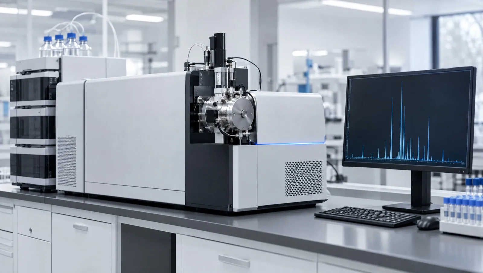
Confirming which cysteines are joined, not merely how many bridges exist, is a separate exercise. The standard approach is to digest the peptide with its disulfides intact, identify the disulfide-linked fragments, then pair them by comparing non-reduced and reduced aliquots of the same digest [11].
- Pepsin suits this well: its acidic pH optimum is a regime in which disulfide rearrangement is precluded, and its broad specificity tends to cleave between half-cystine residues. The cost is complex, overlapping (“ragged”) digests [11].
- Alkaline digestion with trypsin or Lys-C is more specific but creates the artefact it is meant to measure. A 2022 method paper documents scrambled peptides arising during sample preparation at basic pH and elevated temperature, and suppresses them by adding cystamine plus a low concentration of iodoacetamide during denaturation and digestion [13].
- Fragmentation choice and software matter as much as the digest; a 2018 review surveys sample preparation, the fragmentation methods suited to disulfide analysis, and the algorithms that assign linkages from LC-MS/MS data [12].
A quenching step is therefore part of any disulfide method. Lowering the pH is the usual choice; alkylating free thiols (for example with N-ethylmaleimide) is the alternative where the sample must stay near neutral pH [5].
Two structural examples
Insulin. UniProt records the human insulin precursor as a 110-residue chain processed into a B chain of 30 residues and an A chain of 21. Three disulfides are annotated: two between the chains (B7–A7 and B19–A20 in chain numbering) and one within the A chain (A6–A11) [16]. The bridges are what hold the two chains together; without them the molecule is two separate peptides.
Conotoxins. Cone snail peptides are the standard model system for multi-disulfide chemistry. They are small (a median of 26 residues, most between 10 and 45) and are classified into “cysteine frameworks” by the arrangement of their cysteines, with up to five disulfides observed in the family. Framework I peptides (CC-C-C) mostly adopt the globular [1–3, 2–4] connectivity; the ribbon fold is [1–4, 2–3], and a third family forms a knotted arrangement called a cystine knot [3]. One sequence can therefore be assembled into several distinct, isobaric molecules.
What this means in Australia
- Identity includes the bridge pattern. European guidance on synthetic peptides expects the structure of an active substance to state the nature of any cyclisation and the positions involved, and lists mass spectrometry, NMR and peptide mapping among the techniques used to confirm structure [18]. For a cystine-containing research material, an identity test that does not say which disulfide isomer was measured is incomplete.
- Accredited testing. NATA accredits Australian laboratories to ISO/IEC 17025, the international standard for the competence of testing and calibration laboratories [21].
- Chemistry does not change legal status. Several peptides are named in Schedule 4 (prescription only) and Appendix D, clause 5 (possession without authority is illegal) of the current Poisons Standard (October 2026) [20]. The TGA has also stated that a “research use only” disclaimer does not make supply lawful, and does not change a product’s regulatory status, permit importation, or remove advertising or supply obligations [19].
This is general information, not legal advice. Laws change; check the current Poisons Standard and TGA guidance.
Frequently asked questions
What is a disulfide bond in a peptide?
A covalent sulfur–sulfur bond between the side chains of two cysteine residues. The linked pair is called cystine, and it can join two parts of one chain or two separate chains [1, 2, 16].
Why is an oxidised peptide 2 Da lighter?
Because forming the bridge removes one hydrogen atom from each thiol. Reduction adds about 2 Da per disulfide, which is how non-reduced and reduced digests are compared [11].
What are disulfide isomers?
Alternative pairings of the same cysteines. A peptide with three disulfides has 15 possible pairings, all of the same mass, so they are distinguished by chromatography, fragmentation or NMR rather than by mass alone [3].
Why does pH matter so much?
Only the thiolate form reacts, and its concentration depends on pH. Exchange and scrambling accelerate in alkaline solution and slow by orders of magnitude in acid, which is why acidification is used to quench these reactions [5].
Should DTT or TCEP be used to reduce a peptide?
It depends on what follows. DTT is reversible and must be removed before thiol-labelling chemistry; TCEP is effectively irreversible, tolerates some labelling chemistries and resists nickel-catalysed oxidation [5, 10].
How are free thiols measured?
Most often with Ellman’s reagent (DTNB), reading the released TNB at 412 nm against a molar absorption coefficient of about 14,100 M⁻¹ cm⁻¹ at pH 7.3 [5, 7].
References
- National Center for Biotechnology Information. PubChem Compound Summary for CID 5862, L-Cysteine. pubchem.ncbi.nlm.nih.gov/compound/5862. Accessed 25 October 2026.
- National Center for Biotechnology Information. PubChem Compound Summary for CID 67678, L-Cystine. pubchem.ncbi.nlm.nih.gov/compound/67678. Accessed 25 October 2026.
- Akondi KB, Muttenthaler M, Dutertre S, et al. Discovery, synthesis, and structure-activity relationships of conotoxins. Chem Rev. 2014;114(11):5815–5847. doi:10.1021/cr400401e. PMID: 24720541
- Spears RJ, McMahon C, Chudasama V. Cysteine protecting groups: applications in peptide and protein science. Chem Soc Rev. 2021;50(19):11098–11155. doi:10.1039/d1cs00271f. PMID: 34605832
- Winther JR, Thorpe C. Quantification of thiols and disulfides. Biochim Biophys Acta. 2014;1840(2):838–846. doi:10.1016/j.bbagen.2013.03.031. PMID: 23567800
- Nagy P. Kinetics and mechanisms of thiol-disulfide exchange covering direct substitution and thiol oxidation-mediated pathways. Antioxid Redox Signal. 2013;18(13):1623–1641. doi:10.1089/ars.2012.4973. PMID: 23075118
- Ellman GL. Tissue sulfhydryl groups. Arch Biochem Biophys. 1959;82(1):70–77. doi:10.1016/0003-9861(59)90090-6. PMID: 13650640
- Riddles PW, Blakeley RL, Zerner B. Ellman’s reagent: 5,5′-dithiobis(2-nitrobenzoic acid) — a reexamination. Anal Biochem. 1979;94(1):75–81. doi:10.1016/0003-2697(79)90792-9. PMID: 37780
- Cleland WW. Dithiothreitol, a new protective reagent for SH groups. Biochemistry. 1964;3:480–482. doi:10.1021/bi00892a002. PMID: 14192894
- Getz EB, Xiao M, Chakrabarty T, Cooke R, Selvin PR. A comparison between the sulfhydryl reductants tris(2-carboxyethyl)phosphine and dithiothreitol for use in protein biochemistry. Anal Biochem. 1999;273(1):73–80. doi:10.1006/abio.1999.4203. PMID: 10452801
- Gorman JJ, Wallis TP, Pitt JJ. Protein disulfide bond determination by mass spectrometry. Mass Spectrom Rev. 2002;21(3):183–216. doi:10.1002/mas.10025. PMID: 12476442
- Lakbub JC, Shipman JT, Desaire H. Recent mass spectrometry-based techniques and considerations for disulfide bond characterization in proteins. Anal Bioanal Chem. 2018;410(10):2467–2484. doi:10.1007/s00216-017-0772-1. PMID: 29256076
- Nie S, Greer T, Huang X, Zheng X, Li N. Development of a simple non-reduced peptide mapping method that prevents disulfide scrambling of mAbs without affecting tryptic enzyme activity. J Pharm Biomed Anal. 2022;209:114541. doi:10.1016/j.jpba.2021.114541. PMID: 34954467
- Trivedi MV, Laurence JS, Siahaan TJ. The role of thiols and disulfides on protein stability. Curr Protein Pept Sci. 2009;10(6):614–625. doi:10.2174/138920309789630534. PMID: 19538140
- Hawe A, Poole R, Romeijn S, Kasper P, van der Heijden R, Jiskoot W. Towards heat-stable oxytocin formulations: analysis of degradation kinetics and identification of degradation products. Pharm Res. 2009;26(7):1679–1688. doi:10.1007/s11095-009-9878-2. PMID: 19343484
- UniProt Consortium. UniProtKB entry P01308, Insulin (human): chain and disulfide-bond annotations. uniprot.org/uniprotkb/P01308/entry. Accessed 25 October 2026.
- National Center for Biotechnology Information. PubChem Compound Summary for CID 439302, Oxytocin. pubchem.ncbi.nlm.nih.gov/compound/439302. Accessed 25 October 2026.
- European Medicines Agency. Guideline on the development and manufacture of synthetic peptides. EMA/CHMP/CVMP/QWP/367182/2025. Adopted 4 December 2025; effective 1 June 2026. ema.europa.eu. Accessed 25 October 2026.
- Therapeutic Goods Administration. Understanding your responsibilities when importing, compounding and supplying unapproved peptide products. Safety advisory, 13 April 2026. tga.gov.au. Accessed 25 October 2026.
- Therapeutic Goods (Poisons Standard—October 2026) Instrument 2026 (F2026L01327). Federal Register of Legislation. legislation.gov.au/F2026L01327/latest/text. Accessed 25 October 2026.
- National Association of Testing Authorities, Australia (NATA). Accreditation programs, including ISO/IEC 17025 testing and calibration. nata.com.au. Accessed 25 October 2026.