In plain English
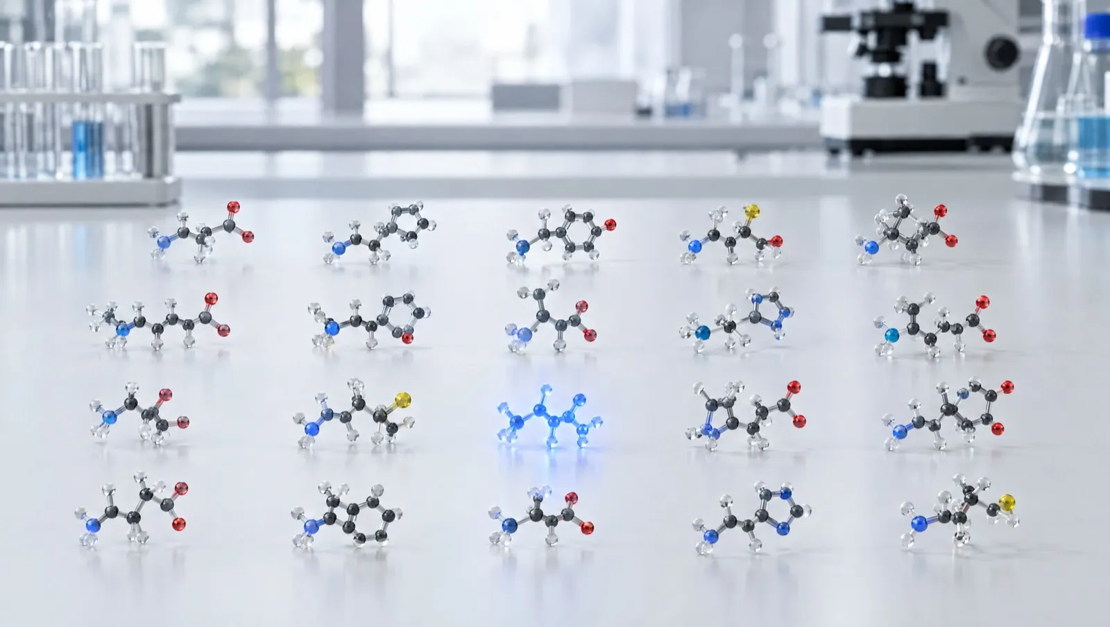
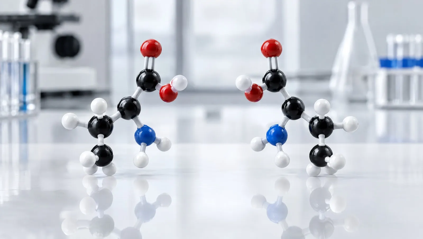
An amino acid has a central carbon with four different groups attached to it. Such a carbon can be built in two ways that are mirror images of each other, like a left and a right hand. Chemists label the two forms L and D [1]. Living cells assemble proteins from the L form only [1]. Glycine is the exception: two of its four attachments are hydrogen atoms, so it has no mirror form [1, 2]. Two standard amino acids, isoleucine and threonine, have a second centre of this kind, so each has four possible arrangements [1, 4, 5].
When peptides are made by chemists, a small share of residues can flip from L to D. This is called racemisation when a mixture of both forms appears, and epimerisation when one centre changes [6, 7]. The flipped molecule has exactly the same mass as the intended one, so measuring the mass cannot reveal it [15]. Finding it needs a method that can tell left-handed from right-handed [7, 16].
The short version
- Chiral amino acids exist as L and D forms, and proteins use L [1].
- Synthesis can flip single residues from L to D [6, 7].
- Flipped forms share the target’s mass, so chiral methods are required [7, 15].
Key facts
| Property | Detail |
|---|---|
| What makes an amino acid chiral | A carbon carrying four different substituents, so the molecule is not superimposable on its mirror image [1] |
| L and D | Prefixes recording configuration at the α-carbon by formal relationship to L- or D-serine [1] |
| Achiral exception | Glycine, whose side chain is a hydrogen atom [1, 2] |
| Two stereocentres | Isoleucine and threonine; their second diastereomers keep the prefix allo [1, 4, 5] |
| Alternative system | The R/S (Cahn–Ingold–Prelog) system [1] |
| L but R | L-cysteine and L-cystine, because sulfur at C-3 outranks the carboxylate [1, 3] |
| Analytical consequence | D/L swaps do not change a peptide’s parent or fragment masses [15] |
What chirality means in an amino acid
The IUPAC-IUB recommendations define the system used in peptide chemistry. Configuration at the α-carbon is designated by the small capital prefix D or L, recording a formal relationship to D- or L-serine and thus to D- or L-glyceraldehyde [1]. The prefix may be omitted when the amino acid is obviously of protein origin and is therefore assumed to be L [1]. An equimolar mixture of both forms is racemic, written DL- or rac- [1].
A second, more general system also exists. In the R/S system of Cahn, Ingold and Prelog, the four substituents are ranked and the centre is R if the first three appear clockwise when viewed from the side away from the lowest-ranked group, and S if anticlockwise [1].
The two systems do not map onto each other neatly. The L configuration of the chiral amino acids found in proteins nearly always corresponds to S, and the recommendations name L-cysteine and L-cystine as the important exceptions, which are R [1]. The reason is a ranking artefact rather than a structural oddity: in most amino acids the priority order around C-2 is NH₃⁺ > COO⁻ > R > H, but in cysteine the sulfur attached to C-3 has a higher atomic number than the oxygen attached to C-1, so the side chain outranks the carboxylate [1]. PubChem accordingly names L-cysteine (2R)-2-amino-3-sulfanylpropanoic acid [3].
Glycine’s side chain is a single hydrogen atom, so its α-carbon carries two identical substituents and has no mirror-image form [1, 2].
Isoleucine and threonine: two stereocentres each
Most standard amino acids have one stereocentre and therefore two forms. Isoleucine and threonine each have a second stereocentre in the side chain, giving four stereoisomers apiece: two enantiomeric pairs, and between the pairs diastereomers (stereoisomers that are not mirror images).
| Amino acid | Configuration | PubChem IUPAC name |
|---|---|---|
| L-isoleucine | (2S,3S) | (2S,3S)-2-amino-3-methylpentanoic acid [4] |
| L-allo-isoleucine | (2S,3R) | (2S,3R)-2-amino-3-methylpentanoic acid |
| L-threonine | (2S,3R) | (2S,3R)-2-amino-3-hydroxybutanoic acid [5] |
| L-allo-threonine | (2S,3S) | (2S,3S)-2-amino-3-hydroxybutanoic acid |
The IUPAC-IUB rules keep the historical prefix allo for exactly these two cases: it should be used only for alloisoleucine and allothreonine, and only with trivial names [1]. For centres other than the α-carbon the rules prefer the R/S system, as in (2S,3R)-threonine [1].
Worked example: how many stereoisomers could a short peptide have?
Oxytocin is a nonapeptide, Cys-Tyr-Ile-Gln-Asn-Cys-Pro-Leu-Gly-NH₂ [18]. Eight of its nine residues have a chiral α-carbon (glycine does not), and its isoleucine contributes a second stereocentre. That is nine stereocentres, so 2⁹ = 512 stereoisomeric sequences are conceivable, exactly one of which is the intended all-L molecule — and all 512 share one molecular formula and one mass.
European guidance makes the same point generally: with the exception of glycine, every natural amino acid in a synthetic peptide carries at least one stereogenic centre, and epimerisation during manufacture means that “in theory, a multitude of different diastereoisomers of the target peptide can be formed” [9].
How racemisation happens during synthesis
A 2023 review of epimerisation in peptide synthesis sets out two general mechanisms, both base-catalysed [6]:
- Oxazolone formation. Strong activation of the backbone carboxylate can form a 5(4H)-oxazolone intermediate; attack by the α-amino group of the same residue or peptide then gives the epimerised product. The review describes this as the predominant source of racemisation during peptide synthesis [6].
- Direct α-proton abstraction. A base removes the α-proton to give a planar anion, and re-protonation from either face gives a mixture of configurations. This matters most where the α-proton is relatively acidic [6].
Several practical factors follow.
| Factor | What the literature reports |
|---|---|
| Carboxyl activation | The activation step itself risks loss of chiral integrity; coupling reagents differ, and additives such as HOBt, Oxyma and Oxyma-B suppress epimerisation [6] |
| Choice of base | Hindered tertiary bases reduce α-proton abstraction; less hindered, more nucleophilic bases increase it [6] |
| Protecting-group type | Derivatives with urethane-type protecting groups (for example Fmoc and Z) are “rather resistant” to oxazolone formation [9] |
| Temperature and heating | In microwave-enhanced Fmoc synthesis of a model 20-mer containing all 20 natural amino acids, cysteine, histidine and aspartic acid were susceptible; lowering the coupling temperature from 80 °C to 50 °C limited racemisation of histidine and cysteine [8] |
| Residue identity | Histidine’s basic imidazole ring makes it prone to epimerisation on activation, and the C-terminal cysteine of a resin-bound peptide is a known weak point whose D-form often co-elutes with the target [6] |
| Cyclisation | Activating a linear precursor’s C-terminus for head-to-tail ring closure is a reported racemisation step [6] |
Two mitigations from the same study are purely procedural: coupling histidine and cysteine conventionally while the rest of the chain is assembled under microwave heating avoided the problem, and the hindered amine collidine minimised formation of D-cysteine [8]. Adding HOBt to the deprotection solution, or using piperazine in place of piperidine, reduced aspartimide formation and the aspartic-acid racemisation that follows it [8]. These side reactions are described from the synthesis side in How Research Peptides Are Made.
Racemisation after synthesis, and during analysis
Configuration is not fixed for ever once the chain is built.
- The succinimide route at Asn and Asp. In a model hexapeptide, L-Val-L-Tyr-L-Pro-L-Asn-Gly-L-Ala deamidated with a half-life of 1.4 days at 37 °C and pH 7.4 to an aspartyl succinimide, which then both hydrolysed (half-time 2.3 h) and racemised (half-time 19.5 h). Most of the racemisation accompanying deamidation was attributed to the rapid racemisation of that intermediate, which is why asparagine and aspartate are described as hot spots for non-enzymatic degradation [10].
- The measurement can create the impurity. Chiral gas chromatography of the constituent amino acids requires acid hydrolysis first, and hydrolysis itself induces some epimerisation. European guidance describes hydrolysing in deuterated hydrochloric acid, so that residues which flip during hydrolysis become deuterated and are distinguished from pre-existing D-residues by a mass spectrometric detector in tandem with the chiral GC [9].
D-amino acids in nature
D-residues are not only laboratory artefacts.
- Bacterial cell walls. D-alanine and D-glutamate are routinely found in bacterial peptidoglycan [11], the sacculus of glycan strands cross-linked by short peptides that forms a closed, bag-shaped layer around the cytoplasmic membrane [12]. Diverse bacteria also make and release non-canonical D-amino acids such as D-methionine and D-leucine, which act as regulators of cell-wall remodelling [11].
- Animal peptides. Many D-amino-acid-containing peptides (DAACPs) have been described in multicellular organisms. A 2014 review groups their origins into incorporation of a D-amino acid, non-enzymatic racemisation, and an enzymatic post-translational step in which a peptidyl aminoacyl L-to-D isomerase converts a residue encoded as L in the mRNA [13].
- Free D-amino acids in mammals. A 2017 overview describes D-serine and D-aspartate in mammalian tissues, with amino-acid racemase in their synthesis and D-amino acid oxidase in their degradation [14].
Why mass spectrometry cannot tell epimers apart
An epimer differs from its parent only in the arrangement of four groups around one carbon. The molecular formula, the monoisotopic mass and the masses of every sequence fragment are identical. As a 2017 analytical paper puts it, standard proteomics methods fail to detect DAACPs because D/L substitutions do not affect the peptide’s parent or fragment masses, and the differences in fragment yields that do exist are often small [15].
Mass spectrometry can still contribute when a separation dimension is added ahead of the mass measurement. That paper reported baseline separation of D/L pairs of up to about 30 residues by trapped ion mobility spectrometry (resolving power up to about 340) followed by time-of-flight mass spectrometry, with linear quantification down to 0.25% of the isomeric mixture; pairs that merged in one charge state were resolved in another [15]. These are specialist methods, not the routine LC-MS identity test described in Reading a Peptide Mass Spectrum.
How D-isomer impurities are actually detected
| Approach | How it works | Notes |
|---|---|---|
| Chiral GC-MS after hydrolysis | The peptide is hydrolysed to free amino acids, separated on a chiral column | Hydrolysis in deuterated HCl distinguishes hydrolysis-induced flips from real ones [9] |
| Marfey’s reagent | Nα-(2,4-dinitro-5-fluorophenyl)-L-alaninamide derivatises the amino acids before HPLC; D- and L-residues become diastereomers that separate on an ordinary reversed-phase column | A review covers the reagent, its analogues, the required precautions and its use for evaluating racemisation [16] |
| Separation of intact diastereomers by RP-HPLC | A single D-substitution often changes retention enough to resolve the diastereomer from the target peptide | Of 35 neuropeptide Y analogues each carrying one D-residue, 28 separated from the native peptide on a C18 column with a triethylammonium phosphate/acetonitrile system; a different stationary phase resolved three more, and capillary zone electrophoresis the remainder [17] |
| LC against epimer standards, or after digestion | Liquid chromatography against reference standards of the likeliest epimer impurities, or chiral analysis of enzymatic digests | Both listed by European guidance alongside chiral GC-MS [9] |
European guidance regards enantiomeric purity as a characterisation test that may not need routine release testing, provided the manufacturer can justify that from a risk analysis or prior knowledge of the epimerisation mechanisms in the process [9]. It adds that control of diastereomers may require specific methods of its own, because one chromatographic method rarely separates every related substance [9]. A 2024 review makes the difficulty explicit: D- and L-isomers have identical physicochemical properties apart from configuration, which is why few validated methods exist [7]. How this appears on a batch report is covered in Common Impurities in Synthetic Peptides, and the deliberate use of D-residues in Peptide Modifications Explained.
What this means in Australia
- Quality expectations. European guidance names enantiomeric and diastereomeric impurities as related substances that a synthetic peptide’s control strategy has to address, both from starting materials and from the process [9]. That is a quality benchmark rather than an Australian legal requirement for research material, but it is the standard against which a chiral purity claim should be read.
- Accredited testing. The National Association of Testing Authorities (NATA) accredits Australian laboratories to ISO/IEC 17025, the international standard for the competence of testing and calibration laboratories [20]. That is a useful signal of method control for a chiral method, which is harder to validate than a routine purity method [7].
- Stereochemistry does not change legal status. Status under the current Poisons Standard (October 2026) attaches to the substance named there; several peptides are listed in Schedule 4 (prescription only) and Appendix D, clause 5 (possession without authority is illegal) [19]. A D-substituted analogue is a different molecule, and whether a class entry captures a specific compound is a legal question.
- Labels do not change status. The TGA has stated that a “research use only” disclaimer does not make supply lawful, and does not change a product’s regulatory status, permit importation, or remove advertising or supply obligations [21].
This is general information, not legal advice. Laws change; check the current Poisons Standard and TGA guidance.
Frequently asked questions
What are D-amino acids?
They are the mirror-image forms of the chiral amino acids. Proteins use L-residues, but D-residues occur in bacterial cell walls, in some animal peptides and as free molecules in mammalian tissues [1, 11, 13, 14].
Why is L-cysteine called R?
Because the R/S system ranks substituents by atomic number. In cysteine the sulfur on C-3 outranks the carboxylate on C-1, which reverses the descriptor even though the configuration is the normal L arrangement [1, 3].
What is the difference between racemisation and epimerisation?
Racemisation produces a mixture of both configurations at a centre. Epimerisation is the conversion of one stereoisomer into another that differs at a single stereocentre [6].
Which residues racemise most during synthesis?
Cysteine and histidine are repeatedly reported as the most susceptible under activation and heating, with aspartic acid affected through aspartimide formation [6, 8].
Can a D-isomer impurity be seen by mass spectrometry?
Not by mass alone, because the epimer has the same parent and fragment masses. It needs a separation step: chiral chromatography after hydrolysis, derivatisation with Marfey’s reagent, reversed-phase separation of the intact diastereomers, or ion mobility ahead of the mass analyser [7, 15, 16, 17].
References
- IUPAC-IUB Joint Commission on Biochemical Nomenclature (JCBN). Nomenclature and symbolism for amino acids and peptides. Recommendations 1983. Biochem J. 1984;219(2):345–373. doi:10.1042/bj2190345. PMID: 6743224. Web version, sections 3AA-3 and 3AA-4: iupac.qmul.ac.uk/AminoAcid. Accessed 25 October 2026.
- National Center for Biotechnology Information. PubChem Compound Summary for CID 750, Glycine. pubchem.ncbi.nlm.nih.gov/compound/750. Accessed 25 October 2026.
- National Center for Biotechnology Information. PubChem Compound Summary for CID 5862, L-Cysteine. pubchem.ncbi.nlm.nih.gov/compound/5862. Accessed 25 October 2026.
- National Center for Biotechnology Information. PubChem Compound Summary for CID 6306, L-Isoleucine. pubchem.ncbi.nlm.nih.gov/compound/6306. Accessed 25 October 2026.
- National Center for Biotechnology Information. PubChem Compound Summary for CID 6288, L-Threonine. pubchem.ncbi.nlm.nih.gov/compound/6288. Accessed 25 October 2026.
- Duengo S, Muhajir MI, Hidayat AT, Musa WJA, Maharani R. Epimerisation in peptide synthesis. Molecules. 2023;28(24):8017. doi:10.3390/molecules28248017. PMID: 38138507
- Badgujar D, Paritala ST, Matre S, Sharma N. Enantiomeric purity of synthetic therapeutic peptides: a review. Chirality. 2024;36(3):e23652. doi:10.1002/chir.23652. PMID: 38448043
- Palasek SA, Cox ZJ, Collins JM. Limiting racemization and aspartimide formation in microwave-enhanced Fmoc solid phase peptide synthesis. J Pept Sci. 2007;13(3):143–148. doi:10.1002/psc.804. PMID: 17121420
- European Medicines Agency. Guideline on the development and manufacture of synthetic peptides. EMA/CHMP/CVMP/QWP/367182/2025. Adopted 4 December 2025; effective 1 June 2026. ema.europa.eu. Accessed 25 October 2026.
- Geiger T, Clarke S. Deamidation, isomerization, and racemization at asparaginyl and aspartyl residues in peptides. Succinimide-linked reactions that contribute to protein degradation. J Biol Chem. 1987;262(2):785–794. PMID: 3805008
- Cava F, Lam H, de Pedro MA, Waldor MK. Emerging knowledge of regulatory roles of D-amino acids in bacteria. Cell Mol Life Sci. 2011;68(5):817–831. doi:10.1007/s00018-010-0571-8. PMID: 21161322
- Vollmer W, Blanot D, de Pedro MA. Peptidoglycan structure and architecture. FEMS Microbiol Rev. 2008;32(2):149–167. doi:10.1111/j.1574-6976.2007.00094.x. PMID: 18194336
- Ollivaux C, Soyez D, Toullec JY. Biogenesis of D-amino acid containing peptides/proteins: where, when and how? J Pept Sci. 2014;20(8):595–612. doi:10.1002/psc.2637. PMID: 24895293
- Genchi G. An overview on D-amino acids. Amino Acids. 2017;49(9):1521–1533. doi:10.1007/s00726-017-2459-5. PMID: 28681245
- Jeanne Dit Fouque K, Garabedian A, Porter J, et al. Fast and effective ion mobility-mass spectrometry separation of D-amino-acid-containing peptides. Anal Chem. 2017;89(21):11787–11794. doi:10.1021/acs.analchem.7b03401. PMID: 28982001
- Bhushan R, Brückner H. Marfey’s reagent for chiral amino acid analysis: a review. Amino Acids. 2004;27(3–4):231–247. doi:10.1007/s00726-004-0118-0. PMID: 15503232
- Kirby DA, Miller CL, Rivier JE. Separation of neuropeptide Y diastereomers by high-performance liquid chromatography and capillary zone electrophoresis. J Chromatogr. 1993;648(1):257–265. doi:10.1016/0021-9673(93)83309-g. PMID: 8245173
- National Center for Biotechnology Information. PubChem Compound Summary for CID 439302, Oxytocin. pubchem.ncbi.nlm.nih.gov/compound/439302. Accessed 25 October 2026.
- Therapeutic Goods (Poisons Standard—October 2026) Instrument 2026 (F2026L01327). Federal Register of Legislation. legislation.gov.au/F2026L01327/latest/text. Accessed 25 October 2026.
- National Association of Testing Authorities, Australia (NATA). Accreditation programs, including ISO/IEC 17025 testing and calibration. nata.com.au. Accessed 25 October 2026.
- Therapeutic Goods Administration. Understanding your responsibilities when importing, compounding and supplying unapproved peptide products. Safety advisory, 13 April 2026. tga.gov.au. Accessed 25 October 2026.