In plain English
pH is a measure of how acidic or alkaline a solution is. Low numbers are acidic, 7 is neutral and high numbers are alkaline. Strictly, pH reflects hydrogen ion activity, which is concentration corrected for how ions behave in a real solution [1].
A buffer is a mixture that keeps pH steady when small amounts of acid or base are added. It contains a weak acid together with its partner base. Each buffer has a number called its pKa, the pH at which the two forms are present in equal amounts. A buffer holds pH best close to its pKa.
pH matters for peptides (short chains of amino acids) because many amino acids carry charges that switch on or off with pH. Those charges change how well a peptide dissolves [15, 16]. pH also speeds up or slows down chemical changes, such as the loss of an amide group from asparagine (deamidation) [17].
The short version
- Choose a buffer whose pKa is within 1 unit of the target pH.
- Some buffers change pH noticeably with temperature [7, 10].
- Buffer pH sets peptide charge and affects stability [15, 17].
- Calibrate the pH meter with two standards that bracket the sample [1].
pH, pKa and the Henderson–Hasselbalch equation
The Henderson–Hasselbalch equation links pH to the ratio of the base and acid forms of a buffer:
pH = pKa + log₁₀ ([base] / [acid])
When the two forms are present in equal amounts, the log term is zero and pH equals pKa. The equation uses concentrations, so it is an approximation; the pH a meter reads depends on activities, which differ from concentrations as ionic strength rises [1].
Worked example: a 50 mM acetate buffer at pH 5.00. Acetic acid has a pKa of 4.76 at 25 °C [2].
- Ratio needed: [acetate] / [acetic acid] = 10^(5.00 − 4.76) = 10^0.24 = 1.74
- Acetate = 50 × 1.74 ÷ 2.74 = 31.7 mM; acetic acid = 50 − 31.7 = 18.3 mM
In practice a laboratory makes up the buffer close to these amounts and then adjusts it to the final pH on a calibrated meter, at the temperature at which it will be used.
Buffering range. At pH = pKa + 1 the base-to-acid ratio is 10:1; at pKa − 1 it is 1:10. Buffer capacity, the ability to absorb added acid or base, is greatest at pH = pKa and falls to about one third of that maximum at pKa ± 1. Outside that window, small additions move pH quickly. This is the reason for the usual rule of choosing a buffer with a pKa within one unit of the target pH.
Common buffers at a glance
| Buffer | pKa near 25 °C | Useful range (approx.) | Temperature sensitivity | Points to watch |
|---|---|---|---|---|
| Acetate | 4.76 [2] | 3.8–5.8 | Low; carboxylic acids change by about ±0.002 per °C [7] | Suits acidic work |
| Phosphate (H₂PO₄⁻/HPO₄²⁻) | 7.09 (pK₂) [3] | 6.1–8.1 | Low near pH 7 [7] | Sodium phosphate can crystallise on freezing and the pH drops [11]; accelerated metal-catalysed oxidation in one model system [19] |
| Tris | 8.07 [4] | 7.1–9.1 | High: about −0.028 pH per °C [7] | Set the pH at the temperature of use |
| Good’s buffers (for example MES, MOPS, HEPES) | Varies by buffer; check a database value | About ±1 around each pKa | Varies by buffer | Many bind metal ions [6]; HEPES with riboflavin and light forms hydrogen peroxide [9] |
The ranges are pKa ± 1. Published pKa values apply to a stated temperature and ionic strength, so they are a starting point, not a final answer [6].
Good’s buffers
In 1966, Norman Good and colleagues described a set of hydrogen ion buffers designed for biological research [5]. Several of them, including MES, MOPS and HEPES, are now standard laboratory reagents [6, 12].
They are not inert. A study of the 20 well-known buffers proposed by Good found that all but three form complexes with metal ions, which can cause serious interference, particularly in protein analyses. MES, MOPS and PIPES were the three non-complexing buffers [6]. Earlier work showed that TES and bicine buffer metal ions as well as hydrogen ions [8]. For metal-binding peptides, such as copper complexes, this matters: a complexing buffer can compete with the peptide for the metal [6, 8].
HEPES has a second known issue. In cell culture medium exposed to visible light, HEPES markedly increased the production of toxic products; the effect required only HEPES and riboflavin with light, and hydrogen peroxide was the main agent [9]. HEPES-containing solutions with riboflavin are therefore best kept away from light.
Temperature: why Tris drifts and phosphate barely moves
Buffers whose acid dissociation releases or absorbs a lot of heat change pKa strongly with temperature. A computer simulation study found that the pH shift with temperature can be substantial for aliphatic amines, which have high enthalpies of acid dissociation, and that the change depends on component concentrations and starting pH [10]. Tris and ethanolamine change by about −0.028 and −0.029 pH units per °C, carboxylic acids by only −0.002 to +0.002 per °C, and phosphate near pH 7 also has a low coefficient [7].
Worked example: Tris made up to pH 8.00 at 25 °C (assuming a constant −0.028 per °C).
- At 4 °C: 8.00 + (0.028 × 21) = about 8.59
- At 37 °C: 8.00 − (0.028 × 12) = about 7.66
The same bottle spans nearly one pH unit between the cold room and an incubator. A Tris buffer is therefore adjusted at the temperature at which it will be used.
Even pharmacopoeial pH standards move. In the Japanese Pharmacopoeia, the phosphate standard reads 6.98 at 0 °C, 6.86 at 25 °C and 6.84 at 40 °C, while the borate standard falls from 9.46 to 9.18 to 9.07 over the same temperatures [1].
Freezing. Freezing can shift pH much more than cooling. In sodium phosphate buffers starting at pH 7.4, crystallisation of disodium phosphate drove the pH of 50 and 100 mM solutions to 4.2 at −10 °C; 8 mM solutions reached 5.2 [11]. Good’s buffers behave differently: HEPES, MOPS and MES became more alkaline on freezing, with MES shifting by up to two units, and blending them with sodium phosphate offset the phosphate’s acidification [12]. Frozen peptide solutions may therefore sit at a different pH from the one written on the label. See Peptide Storage and Stability.
Ionic strength and solvent effects
pH is defined through hydrogen ion activity, the product of concentration and an activity coefficient [1]. Activity coefficients change with ionic strength, so the apparent pKa of a buffer shifts as salt is added or as the buffer itself is made more concentrated. This is why careful pKa measurements state their conditions, for example 25 °C and an ionic strength of 0.10 M [6].
A practical illustration: the Japanese Pharmacopoeia phosphate standard contains equal molar amounts of potassium dihydrogen phosphate and disodium hydrogen phosphate, yet reads pH 6.86 at 25 °C [1], below the pK₂ of 7.09 listed for phosphoric acid [3]. Equal amounts of acid and base forms do not give pH = pKa exactly once ionic strength is taken into account.
Organic co-solvents. Adding acetonitrile, as in HPLC mobile phases, also changes acid–base equilibria. The pKa values of Tris and dihydrogen phosphate have been measured across acetonitrile–water mixtures from 20 to 60 °C, and depend on both solvent composition and temperature [13]. A pH reading taken in water before an organic solvent is added does not describe the mixture.
Ionic strength does not affect every reaction. In one study of small asparagine-containing peptides, deamidation rates were not affected by ionic strength [14].
Peptide charge and the isoelectric point
The isoelectric point (pI) is the pH at which a molecule carries no net charge. For peptides and proteins it depends on the pKa values of the charged groups of seven amino acids and of the terminal amino and carboxyl groups [15]. Below the pI, a peptide carries net positive charge; above it, net negative charge.
Buffer pH therefore decides how charged a peptide is. Solubility is usually lowest near the pI. In a study of protein variants sharing the same hydrophobicity but differing in net charge, each showed a solubility minimum close to its pI, and how steeply solubility fell with pH depended on net charge per residue [16].
Practical consequences:
- A buffer set close to a peptide’s pI invites precipitation or aggregation.
- Moving the buffer pH one or two units away from the pI often improves solubility.
- Sequence-based tools predict pI; one web server reports a prediction error of 0.222 pH units for peptides [15].
Charge counting and solvent choice are covered in Predicting Peptide Solubility; aggregation in Peptide Aggregation.
pH and peptide stability
Deamidation. In a model hexapeptide (Val-Tyr-Pro-Asn-Gly-Ala), deamidation depended markedly on pH, temperature and buffer composition. Between pH 5 and 12 it ran through a cyclic imide intermediate, giving both aspartyl and isoaspartyl products, and buffer catalysis was seen from pH 7 to 11. At acidic pH, direct hydrolysis of the side-chain amide gave only the aspartyl product [17]. The same peptide deamidated with a half-life of only 1.4 days at 37 °C and pH 7.4; replacing the glycine after asparagine with leucine or proline slowed degradation 33- to 50-fold [18]. In other small peptides, isoaspartyl and aspartyl products formed in about a 3:1 ratio at neutral and alkaline pH, and small bases such as ammonia acted as general base catalysts [14]. Buffer species, not only pH, can change the rate.
Methionine oxidation. In small model peptides exposed to an ascorbate and iron(III) system, oxidation of methionine was fastest at pH 6–7. Phosphate accelerated degradation significantly compared with Tris, HEPES and MOPS, and a neighbouring histidine increased the rate further [19].
Cysteine. Thiol reactions generally proceed through the deprotonated thiolate form, so the share of a cysteine present as thiolate, and its reactivity, depend on pH relative to its pKa [20]. More alkaline buffers favour thiolate and therefore thiol reactions, including disulfide formation. See Disulfide Bonds in Peptides.
Buffers do more than set pH. A review of buffers in protein formulations describes how buffers can stabilise proteins through binding or excluded-solute effects, change colloidal stability, and also destabilise them; buffers can degrade themselves [21].
Measuring pH correctly
The Japanese Pharmacopoeia’s pH determination chapter sets out a careful procedure for glass-electrode meters [1]:
- Prepare or buy pH standards and store them in hard glass or polyethylene. Long-stored standards are checked against freshly prepared ones, and alkaline standards are protected from carbon dioxide.
- Condition the electrode by soaking it in water beforehand, and confirm the meter is running stably.
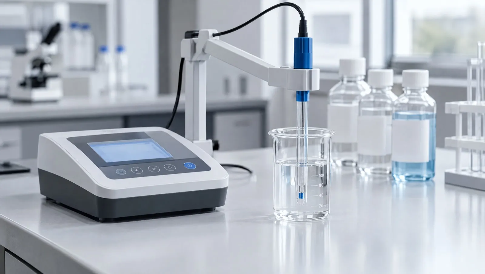
- Calibrate at two points. Standardise first with the phosphate standard, then with a second standard chosen so the expected sample pH lies between the two. Repeat until both read within 0.05 pH unit of their tabulated values without further adjustment.
- Check meters with auto-calibration periodically against two standards, to the same 0.05-unit tolerance.
- Match temperatures. The sample should be within 2 °C of the standards used. The meter’s slope also depends on temperature, from 0.05519 V per pH unit at 5 °C to 0.06610 V at 60 °C.
- Rinse and blot the electrode between solutions, and read only once the value is stable.
- Protect alkaline samples in a covered vessel, under nitrogen if needed. Above pH 11 with alkali metal ions, use an electrode with low alkali error and correct the reading.
The chapter also expects a pH meter to repeat within 0.05 pH unit over five measurements of the same standard [1].
Common errors at the bench
- Calibrating at 25 °C and measuring a cold buffer, or adjusting Tris warm and using it cold [7].
- Using a single standard far from the sample pH.
- Reading pH after adding organic solvent and reporting it as an aqueous value [13].
- Assuming a frozen and thawed buffer still has its original pH [11].
For dilution and molarity arithmetic, see Units and Calculations in the Lab.
Choosing a buffer: a checklist
- Target pH: pick a buffer with a pKa within one unit [2, 3, 4].
- Temperature of use: prefer low-coefficient buffers if the temperature will change, or set the pH at the working temperature [7].
- Freezing: avoid relying on sodium phosphate pH in frozen samples [11, 12].
- Metals: use a non-complexing buffer, such as MES or MOPS, when metal ions matter [6].
- Light: keep HEPES solutions containing riboflavin away from light [9].
- Peptide pI: keep the pH away from the pI to aid solubility [15, 16].
- Stability: consider which pH and buffer species slow deamidation and oxidation for the sequence in hand [17, 19].
What this means in Australia
- Chemistry, not legal status. A peptide’s pI, or the buffer it is dissolved in, has no bearing on how it is regulated. The TGA has stated that a “research use only” disclaimer does not make supply lawful, change regulatory status, permit importation, or remove advertising or supply obligations [22].
- Workplace chemicals. Buffer salts, acids and bases are covered by Australian workplace safety duties. See Safety Data Sheets and Lab Safety.
This is general information, not legal advice. Laws change; check the current Poisons Standard and TGA guidance.
Frequently asked questions
What is a buffer in chemistry?
A mixture of a weak acid and its conjugate base that resists pH change when small amounts of acid or base are added. It works best near its pKa.
What pH range does a buffer cover?
Roughly pKa ± 1. Beyond that, the ratio of base to acid exceeds 10:1 and buffer capacity falls to about a third of its maximum or less.
Why does Tris buffer change pH with temperature?
Tris is an aliphatic amine with a large enthalpy of dissociation, so its pKa falls as temperature rises, by about 0.028 pH units per °C [7, 10].
Why does a phosphate buffer change pH when frozen?
Disodium phosphate can crystallise as the solution freezes, leaving the remaining liquid more acidic; pH 7.4 buffers have reached pH 4.2 at −10 °C [11].
How should a pH meter be calibrated?
With two standards that bracket the expected sample pH, at the same temperature as the sample (within 2 °C), until both read within 0.05 pH unit of their values [1].
Why does pH matter for peptide stability?
pH changes the rate and route of reactions such as asparagine deamidation and methionine oxidation, and the buffer species can catalyse them [17, 19].
References
- The Japanese Pharmacopoeia, 18th edition (English version). General Tests, Processes and Apparatus: 2.54 pH Determination. mhlw.go.jp. Accessed 25 October 2026.
- National Center for Biotechnology Information. PubChem Compound Summary for CID 176, Acetic acid: Dissociation constants. pubchem.ncbi.nlm.nih.gov/compound/176. Accessed 25 October 2026.
- National Center for Biotechnology Information. PubChem Compound Summary for CID 1004, Phosphoric acid: Dissociation constants. pubchem.ncbi.nlm.nih.gov/compound/1004. Accessed 25 October 2026.
- National Center for Biotechnology Information. PubChem Compound Summary for CID 6503, Tris(hydroxymethyl)aminomethane: Dissociation constants. pubchem.ncbi.nlm.nih.gov/compound/6503. Accessed 25 October 2026.
- Good NE, Winget GD, Winter W, et al. Hydrogen ion buffers for biological research. Biochemistry. 1966;5(2):467–477. doi:10.1021/bi00866a011. PMID: 5942950
- Yu Q, Kandegedara A, Xu Y, Rorabacher DB. Avoiding interferences from Good’s buffers: a contiguous series of noncomplexing tertiary amine buffers covering the entire range of pH 3–11. Anal Biochem. 1997;253(1):50–56. doi:10.1006/abio.1997.2349. PMID: 9356141
- Mandaji M, Rübensam G, Hoff RB, et al. Sample stacking in CZE using dynamic thermal junctions I. Analytes with low dpKa/dT crossing a single thermally induced pH junction in a BGE with high dpH/dT. Electrophoresis. 2009;30(9):1501–1509. doi:10.1002/elps.200800584. PMID: 19350541
- Nakon R, Krishnamoorthy CR. Free-metal ion depletion by “Good’s” buffers. Science. 1983;221(4612):749–750. doi:10.1126/science.6879173. PMID: 6879173
- Zigler JS Jr, Lepe-Zuniga JL, Vistica B, Gery I. Analysis of the cytotoxic effects of light-exposed HEPES-containing culture medium. In Vitro Cell Dev Biol. 1985;21(5):282–287. doi:10.1007/BF02620943. PMID: 4019356
- Kipp JE, Schuck DF. Computer simulation of the effect of temperature on pH. J Pharm Sci. 1995;84(11):1347–1352. doi:10.1002/jps.2600841116. PMID: 8587054
- Gómez G, Pikal MJ, Rodríguez-Hornedo N. Effect of initial buffer composition on pH changes during far-from-equilibrium freezing of sodium phosphate buffer solutions. Pharm Res. 2001;18(1):90–97. doi:10.1023/a:1011082911917. PMID: 11336359
- Veselý L, Susrisweta B, Heger D. Making Good’s buffers good for freezing: the acidity changes and their elimination via mixing with sodium phosphate. Int J Pharm. 2021;593:120128. doi:10.1016/j.ijpharm.2020.120128. PMID: 33271311
- Acquaviva A, Tascon M, Padró JM, et al. Automatized sspKa measurements of dihydrogen phosphate and Tris(hydroxymethyl) aminomethane in acetonitrile/water mixtures from 20 to 60°C. Talanta. 2014;127:196–203. doi:10.1016/j.talanta.2014.04.018. PMID: 24913876
- Tyler-Cross R, Schirch V. Effects of amino acid sequence, buffers, and ionic strength on the rate and mechanism of deamidation of asparagine residues in small peptides. J Biol Chem. 1991;266(33):22549–22556. PMID: 1939272
- Kozlowski LP. IPC 2.0: prediction of isoelectric point and pKa dissociation constants. Nucleic Acids Res. 2021;49(W1):W285–W292. doi:10.1093/nar/gkab295. PMID: 33905510
- Tedeschi G, Mangiagalli M, Chmielewska S, et al. Aggregation properties of a disordered protein are tunable by pH and depend on its net charge per residue. Biochim Biophys Acta Gen Subj. 2017;1861(11 Pt A):2543–2550. doi:10.1016/j.bbagen.2017.09.002. PMID: 28890401
- Patel K, Borchardt RT. Chemical pathways of peptide degradation. II. Kinetics of deamidation of an asparaginyl residue in a model hexapeptide. Pharm Res. 1990;7(7):703–711. doi:10.1023/a:1015807303766. PMID: 2395797
- Geiger T, Clarke S. Deamidation, isomerization, and racemization at asparaginyl and aspartyl residues in peptides: succinimide-linked reactions that contribute to protein degradation. J Biol Chem. 1987;262(2):785–794. PMID: 3805008
- Li S, Schöneich C, Wilson GS, Borchardt RT. Chemical pathways of peptide degradation. V. Ascorbic acid promotes rather than inhibits the oxidation of methionine to methionine sulfoxide in small model peptides. Pharm Res. 1993;10(11):1572–1579. doi:10.1023/a:1018960300769. PMID: 8290469
- Ferrer-Sueta G, Manta B, Botti H, et al. Factors affecting protein thiol reactivity and specificity in peroxide reduction. Chem Res Toxicol. 2011;24(4):434–450. doi:10.1021/tx100413v. PMID: 21391663
- Zbacnik TJ, Holcomb RE, Katayama DS, et al. Role of buffers in protein formulations. J Pharm Sci. 2017;106(3):713–733. doi:10.1016/j.xphs.2016.11.014. PMID: 27894967
- Therapeutic Goods Administration. Understanding your responsibilities when importing, compounding and supplying unapproved peptide products. Safety advisory, 13 April 2026. tga.gov.au. Accessed 25 October 2026.